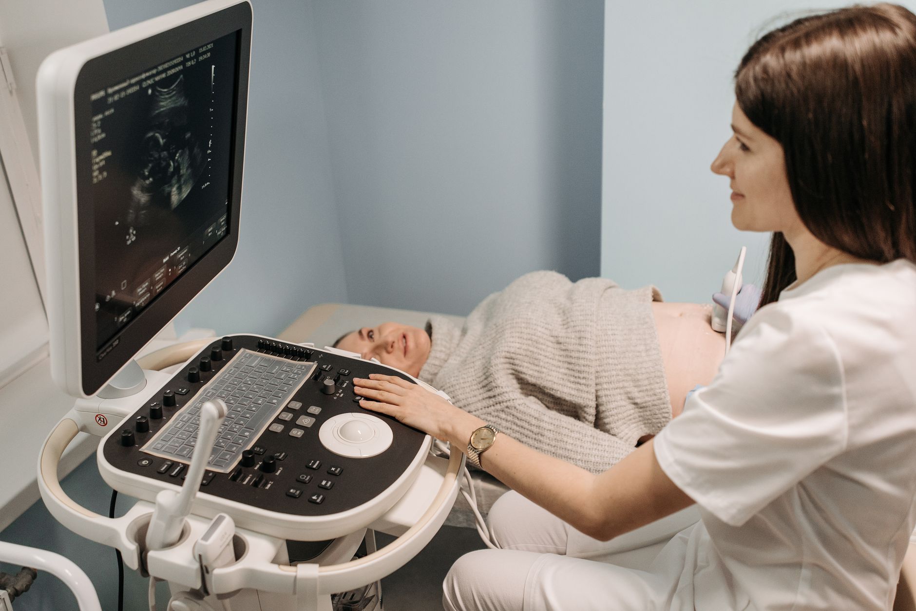
What is a Breast Ultrasound?
A breast ultrasound is a non-invasive medical imaging technique that uses high-frequency sound waves to create detailed pictures of the inside of your breast. It's a crucial diagnostic tool that helps doctors detect and evaluate breast abnormalities, such as lumps, tumors, and cysts. Unlike mammograms, which use X-rays, ultrasounds do not involve radiation, making them a safe option, especially during pregnancy and for younger women or those with dense breast tissue.
Why Might You Need a Breast Ultrasound?
A doctor may recommend a breast ultrasound for several reasons:
- Investigating Abnormalities: If a physical breast exam or a mammogram reveals a suspicious lump, pain, or changes in the breast or nipple area, an ultrasound can help determine the nature of the abnormality.
- Differentiating Cysts from Solid Tumors: Ultrasounds are particularly good at distinguishing between fluid-filled cysts and solid masses. This is important because cysts are usually benign (non-cancerous), while solid masses require further investigation.
- Evaluating Dense Breast Tissue: Women with dense breast tissue may have mammograms that are less effective at detecting small abnormalities. Ultrasound can provide clearer images in such cases.
- For Younger Women and Pregnant Individuals: For women under 25 or those who are pregnant or breastfeeding, ultrasounds are often preferred over mammograms due to the absence of radiation.
- Checking Breast Implants: Ultrasounds can also be used to assess the condition of breast implants and detect any potential issues like leaks.
Types of Breast Ultrasounds
While the basic principle remains the same, there are a few variations of breast ultrasound:
- Standard Breast Ultrasound: This is the most common type, where a sonographer uses a hand-held transducer to scan the breast.
- Automated Breast Ultrasound (ABUS): Also known as AWBUS, this advanced technology uses a larger transducer to capture a 3D image of the entire breast in a single scan. It can be quicker, sometimes taking as little as 5 minutes.
- Contrast-Enhanced Breast Ultrasound (CEBUS): In this method, a contrast agent is injected into a vein. This agent enhances the visibility of blood vessels within abnormalities, helping to provide clearer images and potentially identify cancerous tissues more effectively.
What Happens During a Breast Ultrasound?
The procedure is straightforward and generally painless:
- Preparation: You will be asked to undress from the waist up and lie on an examination table, usually on your back.
- Gel Application: A clear, water-based gel will be applied to your breast. This gel acts as a conductor, allowing the sound waves to travel smoothly between the transducer and your skin.
- Scanning: The sonographer, a trained healthcare professional, will gently move a hand-held device called a transducer over your breast. In some cases, an ABUS machine might be used. The transducer emits sound waves that bounce off the tissues inside your breast, and the echoes are captured to create images on a monitor.
- Image Capture: If the sonographer identifies any suspicious areas, they will capture multiple images for further review by a radiologist.
- Duration: A standard ultrasound might take up to 30 minutes, while an ABUS can be as quick as 5 minutes.
How to Prepare for a Breast Ultrasound
Preparation for a breast ultrasound is simple:
- No Fasting Required: You do not need to fast or avoid eating or drinking before the procedure.
- Avoid Topicals: It's advisable to avoid applying deodorants, powders, lotions, or creams to your chest and underarm area on the day of the test, as these can interfere with the ultrasound waves.
- Comfortable Clothing: Wear comfortable clothing that is easy to remove from the upper body.
Understanding Your Results
After the ultrasound, a radiologist will interpret the images and send a report to your doctor. Your doctor will then discuss the results with you.
- Normal Results: If the ultrasound shows no abnormalities, your doctor will inform you, and no further action may be needed.
- Abnormal Results: If an abnormality is detected, such as a cyst or a solid mass, your doctor will explain what it means. It's important to remember that most breast biopsies, about 4 out of 5, come back negative for cancer. However, further tests might be recommended to confirm the diagnosis.
What Further Tests Might Be Needed?
Depending on the ultrasound findings, your doctor might suggest additional diagnostic steps:
- Biopsy: If a solid mass is detected, a biopsy (taking a small sample of tissue for examination) may be necessary to determine if it is cancerous.
- MRI: In some cases, a breast MRI might be recommended for a more detailed view.
- Follow-up Mammogram: Sometimes, a follow-up mammogram might be advised.
When to Consult a Doctor
It is essential to consult a doctor if you notice any changes in your breasts, such as:
- A new lump or thickening in the breast or underarm area.
- Changes in the size or shape of the breast.
- Changes in the skin of the breast, such as dimpling, puckering, or redness.
- Nipple changes, such as inversion (turning inward) or discharge (fluid leaking from the nipple).
- Persistent breast pain.
Regular breast self-examinations and attending scheduled screening mammograms are vital components of breast health awareness. If you have any concerns, do not hesitate to speak with your healthcare provider.
Prevention and Early Detection
While not all breast conditions can be prevented, early detection significantly improves treatment outcomes. Key strategies include:
- Awareness: Knowing your breasts and what is normal for you is crucial. Report any changes to your doctor promptly.
- Screening: Adhere to recommended screening guidelines, which may include mammograms and ultrasounds as advised by your doctor.
- Healthy Lifestyle: Maintaining a balanced diet, regular physical activity, and limiting alcohol intake can contribute to overall breast health.
A breast ultrasound is a valuable tool in the comprehensive approach to breast health, providing clear images and aiding in the early and accurate diagnosis of various breast conditions. Always consult with your healthcare provider for personalized advice and care.
Additional Medical Guidance
This section adds practical context and preventive advice to help readers make informed healthcare decisions. It is important to verify symptoms early, consult qualified doctors, and avoid self-medication for persistent health issues.
Maintaining healthy routines, following prescribed treatment plans, and attending regular checkups can improve outcomes. If symptoms worsen or red-flag signs appear, immediate medical evaluation is recommended.
Quick Checklist
Track symptoms and duration.
Follow diagnosis and treatment from a licensed practitioner.
Review medication side effects with your doctor.
Seek urgent care for severe warning signs.