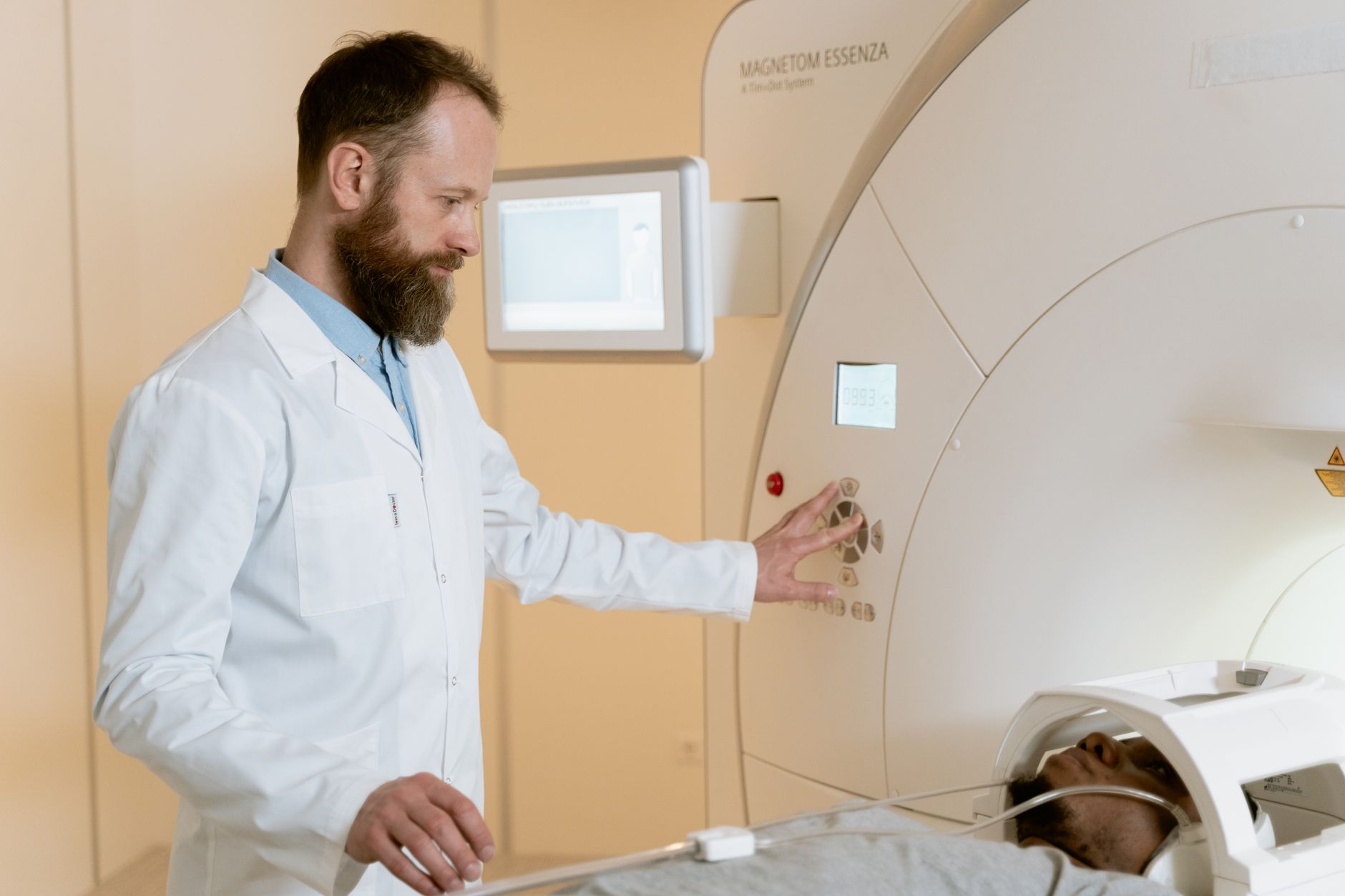
Understanding Magnetic Resonance Enterography (MRE) for Crohn's Disease Crohn's disease is a chronic inflammatory bowel disease (IBD) that can affect any part of the gastrointestinal (GI) tract, from the mouth to the anus. While it most commonly affects the end of the small intestine (ileum) and the beginning of the colon, its impact can be widespread and varied. Diagnosing and monitoring Crohn's disease is crucial for effective management and improving the quality of life for affected individuals. Among the advanced diagnostic tools available, Magnetic Resonance Enterography (MRE) has emerged as a powerful, non-invasive imaging technique that provides detailed insights into the small intestine, making it particularly valuable for evaluating Crohn's disease. What is Magnetic Resonance Enterography (MRE)? Magnetic Resonance Enterography (MRE) is a specialized MRI (Magnetic Resonance Imaging) scan designed to visualize the small intestine. Unlike a standard MRI, MRE uses a combination of strong magnetic fields, radio waves, and a special contrast agent (dye) to create highly detailed, cross-sectional images of the entire small bowel. This allows doctors to meticulously examine the intestinal walls, detect inflammation, identify narrowings (strictures), and spot other abnormalities that are characteristic of Crohn's disease. Why is MRE Used for Crohn's Disease? Crohn's disease is notoriously challenging to diagnose and monitor due to the small intestine's length and its location deep within the abdomen. Traditional methods like endoscopy can only visualize a limited portion of the GI tract. MRE overcomes these limitations by offering a comprehensive view: Accurate Diagnosis: MRE can help confirm a diagnosis of Crohn's disease by clearly showing signs of inflammation, ulcerations, and thickening of the intestinal wall. Assessing Severity and Extent: It helps doctors understand how much of the small intestine is affected and the severity of the inflammation, which is vital for treatment planning. Detecting Complications: MRE is excellent at identifying complications associated with Crohn's disease, such as strictures (narrowed areas that can cause blockages), fistulas (abnormal tunnels connecting the intestine to other organs or the skin), and abscesses (collections of pus). Monitoring Treatment Response: After treatment begins, MRE can be used to assess how well the inflammation is responding to medication, allowing doctors to adjust treatment strategies as needed. Non-Invasive Nature: Unlike surgical procedures or some endoscopic techniques, MRE is non-invasive and does not involve radiation, making it a safer option, especially for patients who may require repeated imaging. How to Prepare for an MRE Test for Crohn's Disease Proper preparation is key to ensuring the best possible imaging results. Your doctor will provide specific instructions, but general guidelines include: Fasting: You will likely be asked to fast for 4–6 hours before the MRE. This helps to clear the digestive tract, allowing for clearer images. Medication Adjustments: Discuss all your current medications with your doctor. Some medications might need to be temporarily stopped or adjusted as they could interfere with the scan. Allergies and Medical History: Inform your doctor about any allergies, especially to contrast agents or iodine, and any previous reactions to imaging tests. Also, disclose any implanted medical devices or metal in your body. Clothing: Wear comfortable, loose-fitting clothing. You will likely be asked to change into a hospital gown to ensure no metal objects interfere with the MRI. Remove all jewelry, watches, belts, hairpins, and any other metal accessories. Intravenous (IV) Line: A small IV line will be inserted into a vein in your arm or hand. This will be used to administer the contrast agent during the procedure. What to Expect During the MRE Procedure The MRE procedure is generally straightforward and painless. Here's what you can expect: Positioning: You will lie down on a comfortable, movable table that slides into the large, tube-shaped MRI machine. Contrast Agent: Before the scan begins, you will drink a special liquid (oral contrast) that helps distend the small intestine and make its lining more visible. This liquid may have a slightly chalky taste. The IV line will be used to inject a gadolinium-based contrast agent, which further enhances the visibility of inflammation and abnormalities. The Scan: The MRI machine uses powerful magnets and radio waves to create detailed images. You will hear loud knocking or buzzing sounds as the machine operates. You may be given earplugs or headphones to help reduce the noise. Staying Still: It is crucial to remain as still as possible during the scan to ensure the images are clear and sharp. Movement can blur the images and affect their diagnostic quality. Breathing Instructions: You might be asked to hold your breath for short periods during the scan. Duration: The MRE scan typically takes about 45 to 60 minutes to complete. Sedation: If you experience claustrophobia (fear of enclosed spaces) or have difficulty staying still, your doctor may offer a mild sedative to help you relax. Recovering from an MRE One of the significant advantages of MRE is its minimal recovery period. In most cases: Immediate Return to Activities: You can usually resume your normal daily activities, including eating and drinking, immediately after the scan. Driving: If you received sedation, you will need someone to drive you home. Avoid driving or operating heavy machinery until the sedative has worn off. Potential Side Effects: Some people might experience mild, temporary side effects such as a metallic taste in their mouth, mild abdominal discomfort, or bloating. These usually resolve on their own within a short period. Contrast Agent: The contrast agent is typically flushed out of your
In summary, timely diagnosis, evidence-based treatment, and prevention-focused care improve long-term health outcomes.