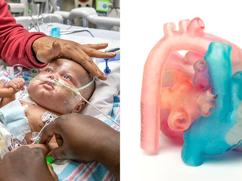
Congenital heart disease covers a wide range of structural problems a baby can be born with, from a small hole between heart chambers to complex defects where the heart's major vessels are arranged in the wrong place entirely. It's one of the most common types of birth defect, and the severity varies enormously from one child to the next.
Here's what makes surgery for these conditions so uniquely difficult. Unlike most adult heart surgery, where anatomy is relatively predictable, no two congenital heart defects look quite the same. A surgeon planning an operation for something like double outlet right ventricle, where both major arteries connect to the wrong chamber, is essentially working out a custom-built puzzle every single time.
Traditional imaging, CT scans, MRI, echocardiograms, gives surgeons flat, two-dimensional slices of a three-dimensional problem. Skilled surgeons learn to mentally reconstruct that 3D picture from years of training, but it's genuinely hard, and mistakes in that mental model can matter enormously once the chest is actually open.
The 2026 Advance: 3D-Printed Hearts You Can Hold
This is where 3D printing has quietly become a real clinical tool rather than a research curiosity. Surgeons now take imaging data from a patient's CT or MRI scan, run it through specialized software to separate out the different heart structures, and print a physical, life-size model of that specific child's heart, valves, chambers, and vessel connections included.
Surgeons can hold the model, rotate it, and in some cases even cut into it before ever touching the actual patient. For a defect as intricate as double outlet right ventricle or an unusual pattern of pulmonary vein connections, being able to physically examine the anatomy beforehand can change decisions made in the operating room.
In clinical practice, this is often the part that gets underappreciated: the value isn't just in the surgeon's understanding, it's in catching a surgical plan that looked reasonable on a flat scan but turns out to be impractical once you can actually see the anatomy in three dimensions. That's the kind of thing a printed model reveals that a screen sometimes doesn't.
Fetal Heart Models: A Newer, More Emotional Use Case
One of the more striking developments has been extending this technology to fetal hearts, meaning models built from ultrasound imaging of a baby's heart before it's even born. Researchers have successfully created 1:1 scale models of fetal hearts, some barely larger than a coin, from prenatal ultrasound data.
These aren't primarily used for surgical planning at that stage, they're used for counseling. A pilot study found that using a printed 3D model during prenatal counseling helped parents understand their unborn child's heart condition at least as well as standard counseling methods, and in some cases seemed to improve their grasp of what was actually going on.
That matters more than it might sound like on paper. Being told your unborn baby has a congenital heart defect is one of the harder conversations in medicine, and a lot of that difficulty comes from parents trying to visualize something abstract from a doctor's verbal description. A physical model that a parent can turn over in their hands changes that conversation in a way I think is genuinely underrated in how we talk about "medical innovation."
Where the Technology Still Has Limits
It would be misleading to present this as a solved problem. Current 3D printing and imaging technology still struggles to accurately capture very thin structures, like heart valves or the wall between the atria, with the same precision as thicker structures like the ventricles. Getting truly valve-accurate models often requires combining several imaging methods together, which isn't universally available.
Cost and access are also real constraints. Not every hospital, especially outside major pediatric cardiac centers, has the imaging software, printers, and trained staff needed to produce these models routinely. For now, this remains concentrated at specialized congenital heart programs rather than being a standard part of every CHD diagnosis pathway.
What This Means for Families Facing a CHD Diagnosis
If your child, or your unborn baby, has been diagnosed with a congenital heart defect, it's reasonable to ask the treating team whether 3D modeling is used at their center, particularly for complex or unusual anatomy. Not every case needs it, simpler defects are often well understood without a physical model, but for the more intricate ones, it can meaningfully shape the surgical approach.
The most important step, though, remains getting care at a center with genuine congenital heart surgery experience. A pediatrician or gynecologist during pregnancy can help direct you toward appropriate fetal cardiology referral if a heart abnormality is suspected on a routine scan.