What is mammogram screening?
A screening mammogram is performed when a person has no obvious breast symptoms and is being checked routinely for possible cancer. A diagnostic mammogram is different. It is generally used when there is a lump, nipple change, abnormal screening result or another specific concern.
That distinction is easy to miss. Doctar's guide to breast cancer in Indian women explains that mammography, ultrasound, MRI and biopsy can have different roles depending on the situation.
Screening is not a guarantee that cancer will be found. Mammograms can miss some cancers, and they can also identify findings that turn out not to be cancer. That is one reason screening decisions involve benefits as well as possible harms.
When should mammogram screening start?
There is no single worldwide schedule that fits every woman. Current guidelines differ.
For example, the U.S. Preventive Services Task Force recommends mammography every two years for women aged 40 to 74 who are at average risk. The American Cancer Society takes a different approach, giving women aged 40 to 44 the option to start yearly screening, recommending annual screening from 45 to 54, and then every two years or yearly screening from 55 onward.
For readers in India, these recommendations should not be treated as a personal prescription. A doctor can consider local guidance, your age, risk factors and previous imaging before recommending a screening plan.
Women with a strong family history, known genetic risk such as BRCA-related risk, previous breast cancer or certain high-risk breast conditions may need a different screening strategy. The USPSTF recommendations for average-risk screening specifically exclude several high-risk groups.
What happens during a mammogram?
The procedure is usually straightforward. You will stand at the mammography machine while a technologist positions one breast at a time between two plates.
The breast is compressed for a few seconds while images are taken. Compression can cause pressure or discomfort, but telling the technologist if you are experiencing significant pain is reasonable.
Doctar's 3D mammogram guide describes the positioning, compression and image-capture process in more detail.
If you are menstruating and your breasts are particularly tender, you may want to discuss scheduling with the imaging centre. The exact timing is not a requirement for everyone, but some people find the examination more comfortable at a less tender point in their cycle.
Is a 3D mammogram better?
Three-dimensional mammography, also called digital breast tomosynthesis, takes multiple X-ray images from different angles and creates a layered view of the breast.
It can provide additional detail and is used widely in breast imaging. The USPSTF considers both digital mammography and digital breast tomosynthesis effective mammographic screening methods.
But “3D” does not automatically mean that every woman needs it. Availability, cost, breast characteristics and the recommendation of the imaging team all matter.
Doctar's 3D mammography article explains how tomosynthesis differs from conventional 2D mammography.
What does breast density mean?
Breast density describes the amount of fibrous and glandular tissue compared with fatty tissue on a mammogram. Dense breasts can make some abnormalities harder to see on a mammogram.
Density also has implications for breast-cancer risk. It does not mean that a person has cancer.
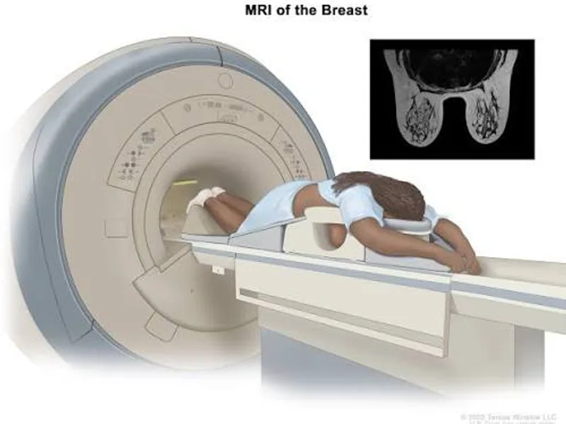
Should everyone with dense breasts automatically have an ultrasound or MRI? Not necessarily. The USPSTF says evidence is currently insufficient to determine the balance of benefits and harms of supplemental ultrasound or MRI after a negative mammogram, regardless of breast density.
Doctar's breast ultrasound information explains that ultrasound can be useful for investigating particular findings and can sometimes complement mammography.
When is breast ultrasound needed?
Ultrasound uses sound waves rather than X-rays. It can help doctors determine whether a finding is fluid-filled or solid and can be useful when investigating a breast lump.
It is often used after an abnormal mammogram or when someone has a specific breast concern. It should not automatically be viewed as a replacement for mammography in routine screening.
Doctar's breast lump guide explains how clinical examination, mammography and ultrasound may work together when a lump is found.
The same principle applies to a movable breast lump: mobility alone cannot establish whether a lump is harmless.
What if the mammogram is abnormal?
An abnormal mammogram does not automatically mean cancer. It means the radiologist has seen something that may need another look.
The next step can include additional mammographic views, ultrasound or, in some cases, MRI. If imaging remains suspicious, a biopsy may be recommended.
Doctar's focal asymmetry guide explains one type of mammographic finding and why additional imaging can sometimes be required.
A breast biopsy report can contain terms such as benign, malignant, grade and hormone-receptor status. These terms should be interpreted with the treating doctor rather than from the report alone.
When does a biopsy become necessary?
A biopsy removes a small sample of tissue so that a pathologist can examine it under a microscope. It is the definitive way to determine whether many suspicious breast findings are cancerous.
One common method is a core needle biopsy. Doctar's core needle biopsy guide explains why doctors may recommend tissue sampling after imaging identifies an area that needs clarification.
The prospect of a biopsy can be frightening. Still, an abnormal mammogram is not the same as a cancer diagnosis, and many abnormal findings have non-cancerous explanations.
Doctar also discusses fat necrosis of the breast, a benign condition that can sometimes resemble a suspicious breast finding on examination or imaging.
Should you get a mammogram if you have symptoms?
Screening is meant for people without signs of breast disease. If you notice a new lump, nipple discharge, skin dimpling, nipple inversion or another persistent change, do not wait for your next routine screening appointment.
Those symptoms require clinical assessment. Depending on your age and the nature of the finding, the doctor may recommend diagnostic mammography, ultrasound or another test.
Doctar's article on breast changes and red spots explains why visible breast changes can have many causes but should be assessed when they persist or look unusual.
A lump below the breast can also have several possible causes. Doctar's guide to a lump around the bra line discusses why examination is more useful than trying to identify the cause at home.
Can men have mammograms?
Yes. Men can develop breast cancer, although it is much less common than breast cancer in women.
Routine mammogram screening is not generally recommended for men at average risk. But a doctor may order breast imaging when a man has a suspicious lump or other breast symptoms.
Doctar's male breast cancer guide explains how examination, mammography, ultrasound and biopsy can be used when a male breast concern needs investigation.
Where can you get a mammogram?
The practical challenge is often finding a suitable imaging centre nearby. Doctar's diagnostic-centre search, for example, lists mammography among available diagnostic tests in Chandigarh.
Similar Doctar listings include mammography testing in Adoni, diagnostic centres in Bhopal, and radiology services in Ramgundam.
Readers can also check diagnostic centres in Ramnagar, pathology and diagnostic services in Belagavi, or ultrasound-centre listings in Godhra.
Doctar also lists radiology centres in Machilipatnam and diagnostic centres in Jalandhar, where mammography appears among the available test categories.
These directories are useful starting points, but availability, equipment, appointment times and reporting arrangements should be confirmed directly with the centre before visiting.
What about hospitals offering breast screening?
Some hospitals combine screening with general surgery, gynaecology or cancer services. For example, LN Medical College and JK Hospital in Bhopal lists breast cancer screening among its services.
In Hyderabad, TX Children's Hospital lists breast cancer screening within its gynaecology and preventive-care services. Patients should verify the age group, imaging modality and appointment process directly with the facility.
For readers who need home-related diagnostic services, Doctar also has home sample collection listings in Surendranagar. Mammography itself generally requires specialised imaging equipment, so home sample collection should not be confused with home mammography.
Does screening work for everyone?
No screening test is perfect. Mammography can detect cancers earlier, but it can also produce false-positive findings, leading to extra imaging or biopsy. Some cancers can also be missed.
The balance between benefit and harm changes with age and individual risk. That is why screening recommendations are written for specific groups rather than as a universal rule.
The American Cancer Society states that screening is intended to find breast cancer before symptoms appear, while its recommendations distinguish average-risk women from those at higher risk.
In clinical practice, this is often missed because the conversation gets reduced to “mammogram every year.” The more useful question is whether the screening schedule matches the person's actual risk.
What should you do before booking?
Have your previous mammogram reports available if possible. Comparing current and older images can help radiologists identify whether a finding is new or unchanged.
Tell the imaging team if you are pregnant, think you may be pregnant, are breastfeeding, have breast implants or have previously had breast surgery. Also mention any new breast symptoms rather than booking the examination as routine screening without explaining them.
If you already have a known high-risk genetic mutation, previous breast cancer or a history of chest radiation at a young age, speak with your doctor about a personalised screening plan rather than following an average-risk schedule.